
Glenohumeral Arthritis Classifications
- Glenoid morphology in OA: Walch classification
- OA with massive rotator cuff tears: Favard classification
- Cuff Tear Arthropathy: Seebauer Classification
- Cuff Tear Arthropathy: Hamada Classification
- Glenoid erosion in cuff tear arthropathy: Sirveaux Classification
- Dislocation arthropathy of the shoulder: Samilson & Prieto Radiological Classification
- Stages of Glenoid wear in RA: Levigne and Franceschi Classification
- Stages of Humeral head wear in RA: Levigne and Franceschi Classification
- Radiological classification of shoulder RA: Levigne and Franceschi Classification
- Radiological classification of RA: Larsen Classification
- Avascular necrosis of humeral head: Neer’s Classification
- Extent of AVN of the Humeral head: Hattrup and Cofield Classification
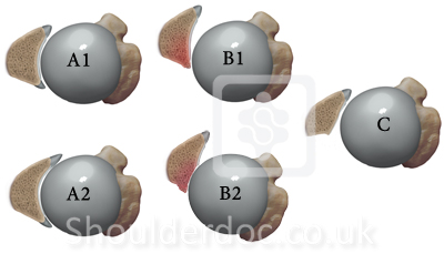
Glenoid morphology in OA: Walch classification
Walch G et al, J Arthroplasty, 14:756-760, 1999
Type A: Humeral head centered
A.1- minor erosion
A.2- major erosion
Type B: Humeral head subluxed posteriorly
B.1- posterior joint space narrow, subchondral sclerosis
and osteophytes
B.2- Retroverted glenoid with posterior rim erosion
Type C: Glenoid retroversion > 25 degrees regardless of erosion
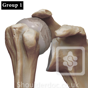
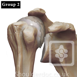
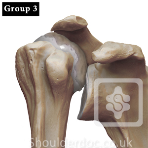
OA with massive rotator cuff tears: Favard classification
Favard et al, OA with massive RCT: the limitations of its current definitions. In: The Cuff, edited by Gazielly D, Elsevier, 1997
Group 1: Humeral head migrated upward,
superior gleno-humeral space narrow,
acromion shaped by humeral head imprint
Group 2: Central gleno-humeral space narrowing,
No change in acromion shape
Group 3: Gleno-humeral joint space narrowing minimal,
Bony destruction / lysis of acromion or humeral head
Cuff Tear Arthropathy: Seebauer Classification
Visotsky, Seebauer et al, JBJS-A, 86-A: 35-40, 2004
Type 1A - Centered stable, Minimal superior migration,
C-A arch acetabularization
Type 1B - Centered medialized, Minimal superior migration,
medial glenoid erosion, C-A arch acetabularization
Type 2 A - Decentered limited stable, superior translation,
superior-medial erosion
significant C-A arch acetabularization
Type 2 B - Decentered unstable, anterior superior escape,
C-A arch and anterior structures deficient
Cuff Tear Arthropathy: Hamada Classification
Hamada et al, CORR, 254: 92-96, 1990
Grade 1: AHI > 6mm
Grade 2: AHI 5mm or less
Grade 3: Grade 2 with acetabularization of acromion
(concave deformity of acromion undersurface)
Grade 4: Grade 3 changes with narrowing of gleno-humeral joint
Grade 5: Bony destruction- humeral head collapse
(AHI: Acromio- Humerus Interval)
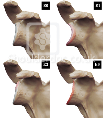
Glenoid erosion in cuff tear arthropathy: Sirveaux Classification
Sirveaux et al, JBJS (B), 86: 388-3985, 2004
E0: Humeral head migration without glenoid erosion
E1: Concentric glenoid erosion
E2: Superior glenoid erosion
E3: Inferior glenoid erosion
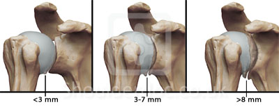
Dislocation arthropathy of the shoulder:
Samilson & Prieto Radiological Classification
Mild Arthrosis: inferior humeral and/or glenoid exostosis < 3mm in height
Moderate Arthrosis: inferior humeral and/or glenoid exostosis
measuring 3mm to 7mm
slight gleno-humeral irregularity
Severe Arthrosis: inferior humeral and/or glenoid exostosis
measuring > 7mm
gleno-humeral joint narrowing and sclerosis
Stages of Glenoid wear in RA: Levigne and Franceschi Classification
Levigne and Franceschi, In: Shoulder Arthroplasty, Edited by Walch and Boileau, 221-230.
Stage 1: Subchondral bone intact or minimally deformed
Stage 2: Erosion reaching the base of coracoid
Stage 3: Erosion going beyond the base of coracoid
Stages of Humeral head wear in RA: Levigne and Franceschi Classification
Levigne and Franceschi, In: Shoulder Arthroplasty, Edited by Walch and Boileau, 221-230.
Stage 1: Subchondral bone intact
Stage 2: Anatomical neck deformed by notch > 10mm
Stage 3: Loss of spherical form of the head
Radiological classification of shoulder RA: Levigne and Franceschi Classification
Levigne and Franceschi, In: Shoulder Arthroplasty, Edited by Walch and Boileau, 221-230.
Ascending form: Most frequent, upward migration of head humerus,
Head retains sphericity, Head initially ascends then medialises,
inferior glenoid notches the humeral neck at late stage
Centered form: Upward migration absent, uniform glenoid wear,
Humeral head pushes into glenoid, progressive head medialisation,
eventual reduction in acromio-humeral distance
Destructive form: Destruction of humeral head, loss of sphericity
notching of humeral neck, simultaneous glenoid destruction
Radiological classification of RA: Larsen Classification
Larsen et al, Acta Radiol Diagn, 18:481-491, 1977
Grade 0: Normal conditions, marginal bone deposits
Grade 1: Slight abnormality, peri-articular soft tissue swelling, osteoporosis or Joint space narrowing
Grade 2: Definite early abnormality, erosion and joint space narrowing present, erosion obligatory except in weight bearing joints
Grade 3: Medium destructive abnormality, erosion and joint space narrowing present, erosion obligatory in all joints
Grade 4: Severe destructive abnormality, erosion and joint space narrowing present, bone deformation in weight bearing joints
Grade 5: Mutilating abnormality, gross bony destruction, dislocation and ankylosis
![]() Top
Top
Avascular necrosis of humeral head: Neer’s Classification
Neer II CS, In : Shoulder Reconstruction, Edited by Neer II CS, 143-271, 1990
Stage 1: Subtle changes, picked up on MRI
Head retains shape, subchondral calcification, pain may be present
Stage 2: Pain present, severe
Articular surface appears intact ,can be indented on pressure
Meniscus sign on radiographs- area of subchondral collapse
Stage 3: Wrinkled/ loose articular cartilage, wedge shape area of subchondral
collapse articular flap, pain ++, step off deformity on x-ray, glenoid normal
Stage 4: Incongruous humeral head, glenoid involvement, secondary arthritis
posterior subluxation, osteochondral loose bodies
Extent of AVN of the Humeral head: Hattrup and Cofield Classification
Hattrup et al, JSES, 8: 559-564, 1999
Group 1: Less than one quarter head involved
Group 2: Between one quarter to one half head involved
Group 3: Between one half and three quarters head involvement
Group 4: More than three quarters head involvement
![]() Top
Top
