
Frontispiece
RUPTURE OF THE SUPRASPINATUS TENDON
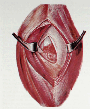
FRONTISPIECE
THANKS to Dr. F. B. Mallory I was able to obtain the autopsy specimen of a case of a completely ruptured supraspinatus, from which this painting was made by Mr. Aitkin.
The skin and subcutaneous tissues were removed; then the fibers of the deltoid separated and held apart by retractors as in the usual routine incision. The diamond-shaped area between the two retractors is the floor of a rather large bursa. Nearly the whole right half of this floor retains its normal, smooth, whitish appearance, but in the left-hand portion of the base or floor is a roughly triangular area which represents the gap formed by the retracted supraspinatus tendon. At the right of this triangular gap, the long head of the biceps appears just beneath the falciform edge of the portion of the musculo-tendi-nous cuff formed by the subscapularis. In the left angle of the triangular area is seen a falciform edge formed by some of the superficial fibers of the infraspinatus. Just superior to this are a few vertical fibers of the deep posterior part of the supraspinatus which have not been evulsed. This was a. very thin, tenuous bit of tissue. The remaining central portion is roughly divided into three parts. The upper, bluish third is the exposed cartilage of the true joint. On its shiny surface near the very edge of the true joint cartilage, we see the high light of the reflection of the window. The lower third of this central space shows a typical "volcano" on the tip of the tuberosity, such as those depicted in Plate V, Figure 1, and in Figures 36 and 40. Between this "volcano" and the cartilage, and also occupying about one-third of the central area and bounded on the right by the margin of the biceps tendon, and on the left by the film-like, untorn edges of the infraspinatus and supraspinatus, we see a red, granulation-like irregular surface. This is the pathologically changed facet of insertion of the supraspinatus tendon and of a portion of that of the infraspinatus from which the tendons have been torn. Compare Figure 40, which is the Rontgen picture of the same specimen.
It must be understood that this picture represents the result of an injury experienced, in all probability, many years before; the tuberosity is in the recessing stage, and the edges of the torn tendons have become smooth by becoming falciform. The distal stub of the supraspinatus tendon, which was probably present in the first few months after the injury, being functionless, has disappeared. The proximal end of the tendon has retracted upward and could only be demonstrated if the newly formed falciform edge of the whole rent were removed. Even in this old case it could be isolated, pulled down and attached to the tuberosity, although with difficulty. One can readily imagine the pain which this patient endured during the first few years after his injury from the mere mechanical irritation from the tuberosity striking on the edge of the acromion during efforts at elevation of the arm, although nature has gradually nearly smoothed off the former prominent tuberosity, and, by partial healing of the edges of the torn structures, has made a new base of approximately spherical surface to pass under the acromion. The writer's operative efforts have mostly been concerned with relieving the results of such conditions. When the general practitioner has learned to recognize the symptoms of these lesions within a few days of their occurrence, suture of such torn tendons will be easily and successfully accomplished.
