
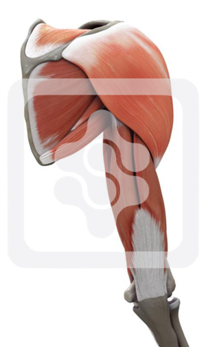
Posterior portal
The figure below shows the muscular anatomy of the right shoulder, as seen from behind. The only constant and useful landmark is the posterior angle of the acromion. The posterior portal is placed 2 cm inferior and medial to this constant point (Figures 1.2 and 1.3).4~7 The first muscle layer that the arthroscope will traverse is the deltoid muscle (Figure 1.4). If the dissection is taken further so that deltoid is detached from the acromion and spine of the scapula, and folded forward (Figure 1.5), the next anatomical layer can be seen.
The first structure to note is the axillary nerve emerging, along with the posterior circumflex humeral vessels, from below teres minor. This neurovascular bundle is only 3 cm below the posterior portal (Figure 1.6), a point of great importance if a second, accessory posterior portal is made in order to perform arthroscopic surgery (for instance, the removal of loose bodies from the infraglenoid recess). The axillary nerve has a singularly inappropriate name, for the first thing it does on leaving the posterior cord of the brachial plexus is to pass below the inferior recess of the shoulder capsule and leave the axilla through the quadrilateral space. As can be seen, this is more a slit than a space, with teres major below, then the long head of triceps medially, humerus laterally, and finally teres minor above it.
As the axillary nerve skirts the inferior border of the shoulder capsule it gives off branches to the joint, and divides into its two terminal branches, the deep and superficial branches. The superficial branch supplies teres minor and then appears behind the posterior border of deltoid to become the upper lateral cutaneous nerve of the arm (not shown on the dissection).
As the axillary nerve skirts the inferior border of the shoulder capsule it gives off branches to the joint, and divides into its two terminal branches, the deep and superficial branches. The superficial branch supplies teres minor and then appears behind the posterior border of deltoid to become the upper lateral cutaneous nerve of the arm (not shown on the dissection).
The head of the humerus has now been osteotomized to expose the anterior capsular structures arthroscopically (Figure 1.10). It is important at this stage to notice the close relationship of the axillary nerve and posterior circumflex artery as they pass directly under the inferior aspect of the joint. The glenoid can clearly be seen along with its central grey spot and surrounding labrum. The long head of biceps can be seen running across the top of the joint cavity, and below it the superior surface of the subscapularis tendon (Figure 1.11).
