
ASD Images
Below are images of a typical ASD (Arthroscopic Subacromial Decompression). The arthroscopic views are inside the subacromial bursa, looking from the back to the front.
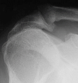
Acromial bone spur seen on x-ray
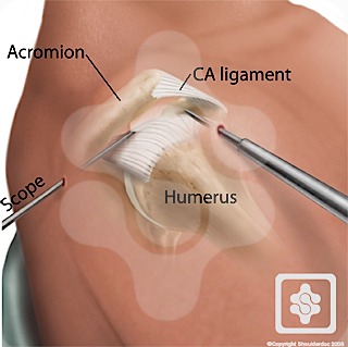
Scope entering bursa from back looking directly at the spur and CA Ligament
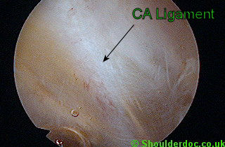
View through scope inside bursa before removal of the CA Ligament
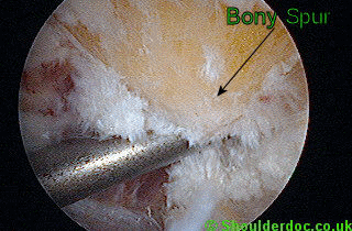
Acromial bone spur seen at arthroscopy, after removal of the CA Ligament.
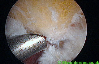
Excising spur with a burr. To appreciate the scale the burr is 4.5mm in diameter, so the spur is about 1.5cm size.
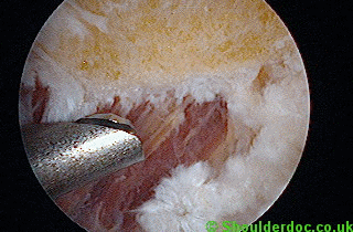
Spur removed. Note the outline of where the spur was originally.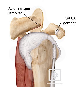
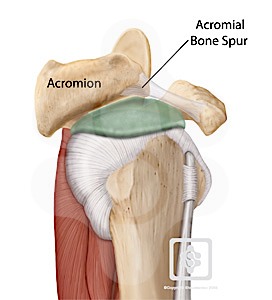
Bone spur removed by surgery with cut ligament
