
Plain radiography
The standard anteroposterior (AP) and axial films of the shoulder will be the first imaging investigation for almost all patients presenting with shoulder disease. To get the most information out of these films, meticulous radiographic technique is essential.
The standard AP projection of the shoulder is taken with the patient slightly oblique, with the arm supinated, and in slight abduction, and should be centred to the coracoid process. The film should be exposed so as to demonstrate both bony and soft tissue detail (Figure 3.1). The acromion, and the greater and lesser tuberosities, should be visible. In the normal patient, the vertical distance between the inferior surface of the acromion, and the superior margin of the humeral head (the so-called acromiohumeral interval) should be at least 7 mm. A reduction in this measurement is suggestive of rotator cuff disease, although it is important to point out that this measurement can be artifactually reduced by radiographic factors, such as centring too low, for example. It is often possible to see a thin, dark line of fat in this region (the peribursal fat plane), which represents extrasynovial fat surrounding the subacromial/subdeltoid bursa. The fat line can be seen as a radiolucency lying deep to the deltoid and extending to the greater tuberosity. It is normally 1-2 mm thick, and is best seen in films taken in slight internal rotation. Failure to visualize the fat plane is a sensitive but nonspecific sign of periarticular disease, including tears of the rotator cuff.1
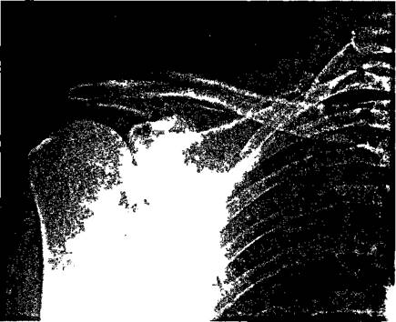
Figure 3.1 Standard AP radiograph of the shoulder.
The AP projection is of great value in the detection of soft tissue calcification (Figure 3.2). Many eponymous views have been described to assist in detecting hydroxyapatite deposition in different components of the rotator cuff. However, unless non-standard views are performed frequently, they are likely to be poorer in quality, and unfamiliar to their interpreters, which limits their value.
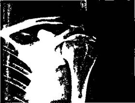
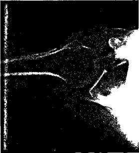
Figure 3.2 Soft tissue calcification on AP radiograph.
Figure 3.3 Standard axial view.
Figure 3.3 shows the standard axial view, which has been taken with the palm down. In this position, the greater tuberosity lies posteriorly, and the lesser anteriorly. In the injured patient, the standard axial view may be difficult to obtain. In these circumstances, a modified axial view, developed by Wallace and colleagues 2 can be helpful. In this technique, the patient is sitting rotated, so that the scapula is parallel to the edge of the horizontal cassette, which is in contact with the arm. The tube is angled at 30 degrees. In this position, the central ray passes just lateral to the coracoid process, through the joint, to the cassette. The effect of this is to produce a magnified, and slightly distorted image of the shoulder (Figure 3.4), but with the advantage that the patient can be X-rayed without removing slings or collars and cuffs.
The search for the hatchet lesion, the humeral head defect produced by recurrent anterior dislocation, has generated a large number of eponymous radiographic projections. Whenihe lesion is large, it can be demonstrated by an internally rotated AP projection, or a palm-down axial. More subtle lesions may require specialized techniques like the Stryker view, arthrography, or CT to demonstrate them.
A number of indirect plain film signs of rotator cuff disease have been described. In 1964, Cotton and Rideout3 analysed over 100 cases, both radiologically and pathologically, at postmortem. They found that cyst formation in the upper two-thirds of the anatomical neck was a reliable and constant sign. The acromiohumeral interval was reduced in many cuff tears, but could be normal in the presence of severe disruption. Sclerosis of the greater tuberosity was a very unreliable sign, but sclerosis or remodelling of the inferior acromial surface correlated well with the presence of a cuff tear. The formation of a subacromial spur, an area of new bone formation arising from the inferior surface of the acromion4 has also been described in some patients with full thickness tears.5
In general, plain films of the shoulder are of greatest value in acute trauma, and in the assessment of calcific periarthritis. Many abnormalities can be seen which suggest the presence of soft tissue disease, but these are usually non-specific, and the X-ray may be completely normal in the presence of a complete rotator cuff disruption.

Figure 3.4 Nottingham axial view.
