
Arthroscopic Subacromial Decompression
Impingement
Our understanding of the impingement syndrome has greatly improved over the past few years and there is no doubt that this will continue for the next decade. It is becoming apparent that impingement is just the final common pathway for a number of different pathologies. Perhaps the best way to look at impingement is anatomically and functionally.
The subacromial space is not a true space but a potential space. Thus it has a roof and a floor, but no real walls. This potential space is lined by the subacromial bursa. The subacromial space is roofed by the acromion, the acromioclavicular joint, the extreme distal portion of the clavicle, the coracoacromial ligament and, anteriorly, the coracoid process itself. The floor of the space is the rotator cuff, the coracohumeral ligament and the long head of biceps. Once distended, the potential space becomes a space: the anterior wall consists of the coracoid process, the lateral and posterior walls are formed by the deltoid and the medial wall is made of bands from the coracoacromial ligament to the acromioclavicular joint and the musculotendinous junction of supraspinatus (Figure 8.1).
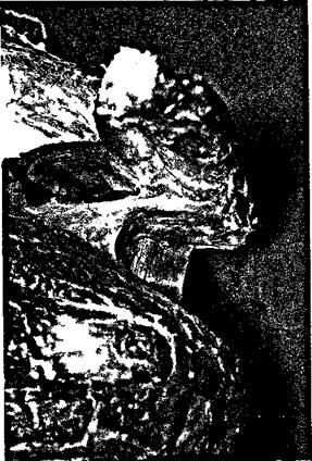
Figure 8.1 Anatomy of the subacromial space.
The subacromial space can therefore be compromised either by the roof coming down, or the floor coming up. The roof can only come down by some part of it being thicker anatomically, but the floor can come up both anatomically (by a thickening of the floor), or functionally (by upward subluxation of the glenohumeral joint, which can be physiological or pathological).
A 'lower' roof
The acromion
The shape of the anterior acromion varies from person to person, and may be flat or hooked. The Bigliani classification recognizes three types of shape, varying from flat (type 1) to fully hooked (type 3). The best way to appreciate the acromial shape is to take a subacromial arch radiograph. The relevance of the Bigliani type 3 acromion is that the hook will have to be surgically excised.
The acromioclavicular joint
Figures vary but, on average, 30 per cent of patients with impingement will have degenerate changes visible in the acromioclavicular joint on an anteroposterior radiograph of the shoulder. A proportion of these will have inferior osteophytes (Figure 8.2) projecting down into the subacromial space. The capsule of the acromioclavicular joint may also be thickened. Obviously, to remove the osteophytes, the capsule has to be resected first.
Figure 8.2 Inferior osteophytes projecting down from the acromioclavicular joint.
The coracoacromial ligament
We have already shown that the coracoacromial ligament inserts underneath the acromion and not onto the front of the acromion, so any thickening of this ligament at its insertion point will project into the subacromial space.
The coracoid process
Gerber et al[1] have described a variety of anterior impingement caused by the proximity of the coracoid to the front of the shoulder, which is relieved by the operation of coraco-plasty.
A 'higher' floor (functional)
Physiological
Matthews and Fadale[2] have shown how the subacromial space is diminished with abduction of the arm. Sigholm et al,[3] using a microca-pillary infusion technique, have measured the pressure in the subacromial space. At rest, the pressure was 8mmHg, rising to 32mmHg with the arm abducted to 45 degrees and to 56 mmHg with the arm abducted to 45 degrees while holding a 1 kg weight. This 'physiological impingement' has relevance both to work-related and sport-related impingement.
Pathological
Upward subluxation of the glenohumeral joint may be caused by instability, or by an abnormality of muscular or nervous control of the rotator cuff. In particular, rotator cuff tears can lead to upward subluxation (Figure 8.3). Weakness of the rotator cuff, secondary to suprascapular nerve entrapment, may cause upward subluxation.
.jpg)
Figure 8.3 Upward subluxation of the humeral head following rotator cuff tear.
Obviously an understanding of the aetiology of impingement is vital to the correct management. For instance, if a particular patient has impingement secondary to instability, then the capsule should be repaired. Subacromial decompression will only make this patient worse. A patient with anterior impingement between the cuff and the coracoid needs a coracoplasty and will not get better with a subacromial decompression. A patient with work-related physiological impingement may need only to change his posture, for example, to working with the elbow at the side instead of abducted. Patient selection is the key to success with subacromial decompression.4
Natural history of impingement
Neer[5] classified the impingement syndrome into three stages according to changes in the rotator cuff, caused by repeated abrasion against the roof of the subacromial space. Stage 1 causes oedema and haemorrhage in the cuff, occurs in the younger patient, is often work- or sport-related and tends to settle with conservative measures. Stage 2 causes fibrosis and 'tendinitis' or partial thickness tears, occurs in an older age group and is more resistant to therapy. Stage 3 implies full thickness rotator cuff tears, rupture of the biceps tendon and bone changes.
Chard et al[6] reviewed 137 patients with impingement treated conservatively. At a mean of over 18 months, 35 still had active tendinitis, a further 40 still had residual pain and 8 had developed pain due to other causes. Only 54 resolved and these were distinguished by early presentation and a history of overuse unrelated to occupation. In 29 patients, function was still impaired and 2 lost their jobs. Chard and colleagues concluded that rotator cuff tendinitis is not an early self-limiting condition, that a sizeable proportion did not resolve with conservative management and that improvements in management were needed.
