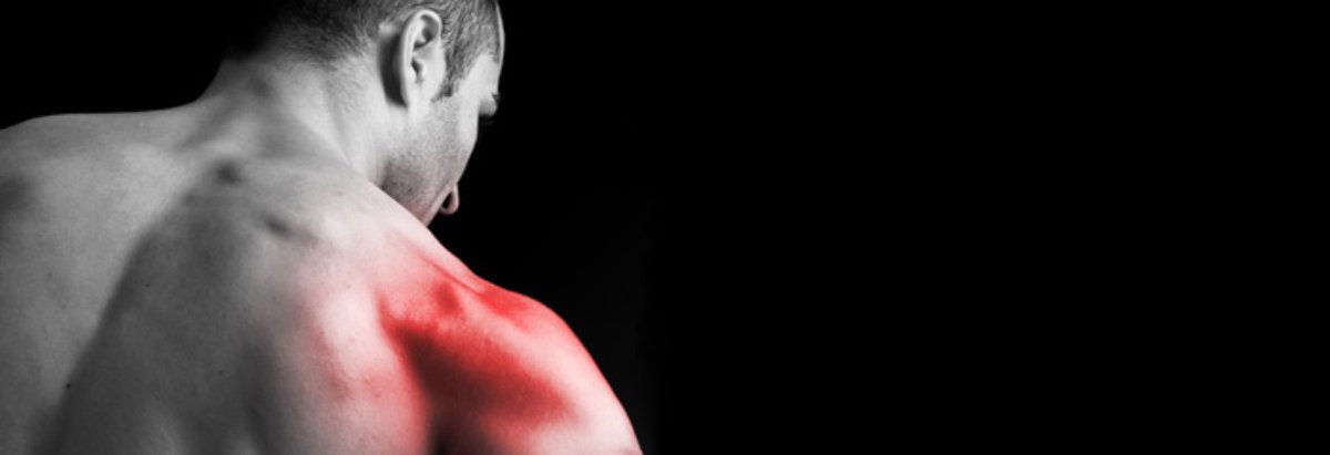
Posterior Portal

Figure 1.1 shows the muscular anatomy of the right shoulder, as seen from behind. The only constant and useful landmark is the posterior angle of the acromion. The posterior portal is placed 2 cm inferior and medial to this constant point (Figures 1.2 and 1.3).4~7 The first muscle layer that the arthroscope will traverse is the deltoid muscle (Figure 1.4). If the dissection is taken further so that deltoid is detached from the acromion and spine of the scapula, and folded forward (Figure 1.5), the next anatomical layer can be seen.

The first structure to note is the axillary nerve emerging, along with the posterior circumflex humeral vessels, from below teres minor. This neurovascular bundle is only 3 cm below the posterior portal (Figure 1.6), a point of great importance if a second, accessory posterior portal is made in order to perform arthroscopic surgery (for instance, the removal of loose bodies from the infraglenoid recess). The axil­lary nerve has a singularly inappropriate name, for the first thing it does on leaving the posterior cord of the brachial plexus is to pass below the inferior recess of the shoulder capsule and leave the axilla through the quadrilateral space. As can be seen, this is more a slit than a space, with teres major below, then the long head of triceps medially, humerus laterally, and finally teres minor above it.

SHOULDER ARTHROSCOPY


Figure 1.1 Muscular anatomy of the right shoulder seen from behind. The constant landmark is the posterior angle of the acromion, which you can feel on your own shoulder.

Figure 1.2 The posterior portal is situated 2 cm medial to and 2 cm inferior to the posterior angle of the acromion, as marked on this patient.

As the axillary nerve skirts the inferior border of the shoulder capsule it gives off branches to the joint, and divides into its two terminal branches, the deep and superficial branches. The superficial branch supplies teres minor and then appears behind the posterior border of deltoid to become the upper lateral cutaneous nerve of the arm (not shown on the dissection).

Figure 1.8 In this dissection, the spine of the acromion has been osteotomized and removed along with trapezius and deltoid. Supraspinatus and infraspinatus have then been lifted from their origins on the blade of the scapula to show the suprascapular nerve passing around the edge of the spine to supply both muscles.

Figure 1.9 Infraspinatus and teres minor have now been reflected back and the posterior capsule of the shoulder has been incised at its origin from the neck of the glenoid and reflected laterally. The 'bare area' of the humeral head can be seen.

Figure 1.10 The head of the humerus has now been osteotomized and removed exposing the glenoid, the glenoid labrum, the long head of biceps and the anterior glenohumeral ligaments.

Figure 1.11 A simplified interpretation of the capsular structures as seen at arthroscopy.

The head of the humerus has now been osteotomized to expose the anterior capsular struc­tures arthroscopically (Figure 1.10). It is impor­tant at this stage to notice the close relationship of the axillary nerve and posterior circumflex artery as they pass directly under the inferior aspect of the joint. The glenoid can clearly be seen along with its central grey spot and surrounding labrum. The long head of biceps can be seen running across the top of the joint cavity, and below it the superior surface of the subscapularis tendon (Figure 1.11).

This website is certified by Health On the Net Foundation. Click to verify. This site complies with the HONcode standard for trustworthy health information:
verify here.

ShoulderDoc.co.uk satisfies the INTUTE criteria for quality and has been awarded 'editor's choice'.

The material on this website is designed to support, not replace, the relationship that exists between ourselves and our patients. Full Disclaimer