Cannulation
If the instruments are passed freehand, it is best to make a track for them first, by inserting a sharp trochar and cannula down the anterior portal. However, this does not necessarily guarantee that the instruments will follow the track and, if repeated passages are envisaged, then it is best to insert a cannula. There are two methods to pass a cannula, the outside-in technique or the inside-out technique.


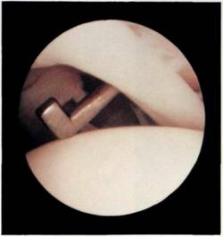
Figure 4.28 A hook probe is then inserted through the anterior portal, using either the inside-out or outside-in technique.
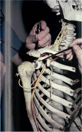
Figure 4.29 The superior portal can be used for irrigation.
Outside-in technique
The arthroscope is brought into the foramen of Weitbrecht and rests on the synovium just above the subscapularis tendon. In the thin patient, the arthroscope can be felt tenting up the soft tissues. Alternatively, the room lights can be lowered, which makes light from the arthroscope visible through the skin, if thin enough. Having marked this site as the insertion site, a 5mm skin incision is made and the arthroscope backed off so that the plunging sharp cannula does not damage it. The trochar and canula are inserted, aiming at the site from which the arthroscope tip has been withdrawn. The sharp trocar and cannula are carefully advanced with rotaion until joint entry occurs.
Inside-out technique
The inside-out technique is more elegant and was devised by Dr A. Wissinger. The arthroscope is advanced through the foramen of Weitbrecht above the subscapularis tendon, as before, until it rests on the synovium. In this position, the arthroscope is removed, leaving the arthroscope cannula resting on the synovium at the front of the shoulder. A long sharp rod (Wissinger rod) is now passed down the arthroscope cannula from the back of the shoulder and pushed out of the front of the shoulder. As it tents the skin, a 7 mm stab incision is made over it. A cannula is then placed over the rod in front of the shoulder and 'railroaded' down the rod and into the shoulder joint. With both cannulae inside the shoulder joint, one from the posterior portal and the other from the anterior portal, the Wissinger rod is withdrawn and the arthroscope reinserted.
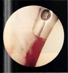
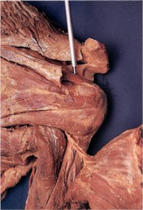
An additional portal can now be used for irrigation (Figure 4.29), the superior or Neviaser portal (see Chapter 3). A Verres needle is placed medial to the acromion in the suprascapular fossa and triangulated to enter the superior part of the joint (Figure 4.30). Some concern has been expressed10 that this portal may damage the rotator cuff but, in dissections at Nottingham and at other centres, it has been seen that the needle passes through the muscle of supraspinatus and not the tendon (Figure 4.31). The needle can be tucked down in the posterior gutter to avoid instrument clashes. Most procedures can be performed with a two-portal technique, and this third portal is rarely needed.


Figure 4.30 The Verres needle can be used to suck blood from the joint.
Figure 4.31 The needle passes through muscle and not through tendon.


